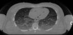
CT projection data for assignments
The data was obtained was obtained from a female patient using a Toshiba
scanner at Rigshospitalet. More information can be found in the DICOM files described below.
The data for the measurements are given below:
| Scanner: |
Toshiba |
| Number of projections: |
200 |
| Samples for each projection: |
512 |
| Image size: |
512 x 512 |
| Projection: |
Parallel beams |
The data can be obtained from the ftp-server. Several files are found. The properties are listed in the following
table (click on the file name to get the data):
The files should be downloaded in binary format. The files can be directly
read and manipulate with Matlab. The variable ct_data contains the projection
data with 200 angular projections and 512 values for each projection.
The pixel values are equal to Hounsfield units and a pixel value of
zero corresponds to -1000 in Hounsfield units. The projected data are also off-set
1000 Hounsfield units.
More information about the images can be found in the DICOM files located in
https://courses.healthtech.dtu.dk/22485/files/ct_data/dicom_data/dicom_data_zip.zip
The individual files can be read by the DICOM reader functions in Matlab. An example is given
in the script:
https://courses.healthtech.dtu.dk/22485/files/ct_data/dicom_data/display_dicom_images.m,
which load all images and shows a video with them. The whole file structure can be downloaded
using the zip-file:
https://courses.healthtech.dtu.dk/22485/files/ct_data/dicom_data/dicom_data_zip.zip. Note that
it is big - 193 MBytes.
| 

 Print Version